3D Diagnostic Detail
Visualize bone volume, nerve canals, sinus floors, and tooth roots in three dimensions. This level of detail helps us identify pathology and plan procedures with confidence that 2D images can’t provide.
3D X-rays that enhance diagnosis and surgical planning.
Cone-beam computed tomography (CBCT) produces a detailed 3D X-ray of your teeth, jawbone, nerves, sinuses, and airway — all in a single, quick scan. Unlike traditional 2D X-rays that flatten anatomy into a single plane, CBCT lets us rotate, slice, and measure structures from every angle, revealing information that would otherwise require referral to a hospital CT.
We use CBCT selectively — only when the clinical question demands 3D detail that standard X-rays cannot answer. Common scenarios include implant planning, evaluating impacted teeth, diagnosing jaw pathology, assessing root fractures, and evaluating the temporomandibular joint (TMJ) and airway for sleep-related concerns.
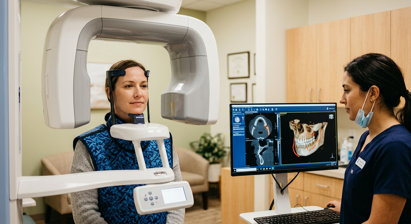
How CBCT works: You stand or sit while a compact arm rotates around your head in about 10–20 seconds. The machine captures hundreds of individual X-ray projections, and software reconstructs them into a high-resolution 3D volume. We can then navigate through cross-sections in any plane — axial, sagittal, or coronal — and take precise measurements of bone height, width, density, and nerve-canal proximity.
What CBCT helps us diagnose and plan: implant site evaluation (bone volume, sinus proximity, nerve mapping), impacted or supernumerary teeth, jaw cysts or tumors, root fractures and resorption, TMJ disorders, airway assessment for sleep apnea, and guided surgical planning for complex extractions or grafting procedures.
Visualize bone volume, nerve canals, sinus floors, and tooth roots in three dimensions. This level of detail helps us identify pathology and plan procedures with confidence that 2D images can’t provide.
Virtually place implants, measure bone density, and map nerves before any incision. Guided surgery templates created from CBCT data improve safety and accuracy.
Dental CBCT uses a focused, cone-shaped beam that limits radiation to the area of interest. Typical doses are a fraction of a full medical CT scan, and we only prescribe it when the diagnostic benefit clearly justifies the exposure.
We position you comfortably — usually standing or seated with a chin rest. The scan arm rotates around your head for 10–20 seconds while you hold still.
Software reconstructs the scan into a 3D volume within minutes. We review cross-sections, panoramic views, and 3D renderings together on screen.
Measurements and annotations guide treatment planning — whether for implants, extractions, grafts, orthodontics, or airway evaluation.
If guided surgery is planned, we design a virtual surgical guide from your CBCT data, which is then 3D-printed for precise implant placement.
How much radiation does a CBCT scan involve?
Doses vary by field of view and machine settings, but a typical dental CBCT delivers roughly 5–20% of a standard medical CT of the same area. We follow ALARA (As Low As Reasonably Achievable) principles and only prescribe CBCT when 3D information will change or improve your care.
How long does the scan take?
The actual exposure is usually 10–20 seconds. Including positioning and review, plan for about 5 minutes total.
Will I feel claustrophobic?
Unlikely. Unlike a medical MRI or CT tunnel, dental CBCT is an open design — the arm rotates around you while you stand or sit in the open. Most patients find it very comfortable.
Can I have a CBCT scan if I’m pregnant?
We generally avoid non-urgent radiographic imaging during pregnancy. Please let us know if you are or may be pregnant so we can weigh the clinical need carefully.
Is CBCT covered by dental insurance?
Coverage varies by plan and clinical indication. Many plans cover CBCT for implant planning or pathology evaluation. We verify benefits and provide an estimate before scheduling.
How is CBCT different from a regular dental X-ray?
Traditional dental X-rays produce a 2D image, which can overlap structures and hide detail. CBCT produces a full 3D volume you can slice in any direction, revealing bone depth, nerve positions, and anatomy that flat images miss.
Do I need CBCT for every implant?
Not always, but for most implant cases, CBCT provides critical information about bone volume, density, and proximity to nerves and sinuses. It’s considered the standard of care for implant planning by most oral surgery and implant guidelines.
Can CBCT detect infections or tumors?
Yes. The 3D detail can reveal periapical pathology, cysts, and certain tumors that may not be visible on 2D X-rays. Early detection through CBCT can lead to more timely and effective treatment.