Crystal-Clear Detail
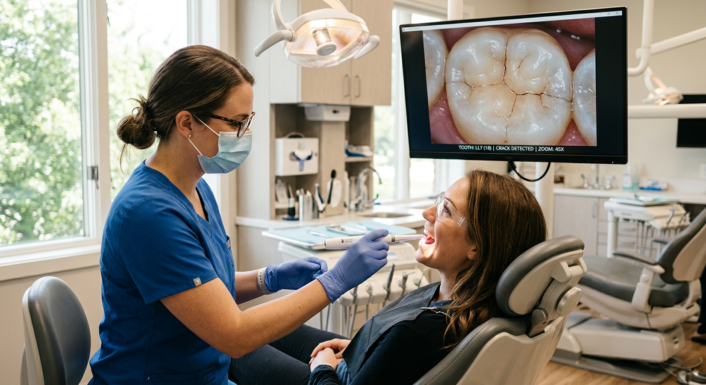
See your teeth at up to 100x magnification on a full-size monitor. Cracks, wear, and early decay that are invisible to the unaided eye become clearly visible, helping us catch problems early.
High-definition images that help you see what we see.
An intraoral camera is a pen-sized, high-definition camera that captures magnified images of your teeth, gums, and soft tissues. By illuminating and photographing areas that are difficult to see with the naked eye — or even with a dental mirror — the camera reveals cracks, early decay, worn restorations, plaque buildup, and tissue changes in vivid detail.
We display these images on a chairside monitor so you see exactly what we see. This transparency transforms the conversation about your dental health: instead of taking our word for a finding, you can view the evidence yourself, ask questions, and make confident, informed decisions about treatment.
How the intraoral camera works: The camera head is about the size of a dental mirror. It uses focused LED lighting and a high-resolution sensor to capture still images and, in some cases, live video. Images are instantly displayed on a nearby monitor at up to 100x magnification. We can freeze, zoom, annotate, and save any frame to your digital chart for future comparison.
What the intraoral camera helps us show you: hairline cracks in enamel, early-stage cavities, chipped or worn fillings and crowns, gum recession and tissue inflammation, plaque and calculus deposits, broken or leaking sealants, and before-and-after comparisons for completed treatments. These images also help us communicate more clearly with dental labs and specialists.
See your teeth at up to 100x magnification on a full-size monitor. Cracks, wear, and early decay that are invisible to the unaided eye become clearly visible, helping us catch problems early.
There’s no guesswork — you see exactly what we see. This visual evidence helps you understand why treatment is recommended and makes shared decision-making natural.
We save every image in your digital chart for side-by-side comparison at future visits. This makes it easy to monitor a watch area, evaluate a restoration, or celebrate the results of completed treatment.
During your exam or cleaning, we gently glide the camera tip along your teeth and gums, capturing close-up images of each area.
Images appear instantly on the chairside monitor. We pause to discuss anything noteworthy — a crack, stain, worn filling, or healthy tissue.
Selected images are annotated and saved to your digital chart. If needed, we share them with specialists or labs for treatment coordination.
At follow-up visits, we compare new images to previous ones to track changes, verify healing, and confirm the success of completed treatment.
Does the camera hurt?
Not at all. The camera tip is small, smooth, and used gently — similar to a dental mirror. There’s no radiation, pressure, or discomfort involved.
Is there any radiation?
No. The intraoral camera uses safe, visible LED light — not X-rays. It’s completely radiation-free and safe for everyone, including pregnant patients and children.
Will I be able to see the images?
Absolutely — that’s the whole point. We display images on a monitor right next to you so you can follow along with the exam in real time.
Can I get copies of my photos?
Yes. We’re happy to provide copies of your images for your personal records, and we can share them electronically with other dental providers if you need a referral.
How often are intraoral photos taken?
We typically capture images during comprehensive exams, when monitoring a specific concern, or as part of treatment planning for cosmetic or restorative work. Frequency depends on your individual needs.
Does this replace X-rays?
No. Intraoral cameras photograph surfaces and external detail, while X-rays reveal internal structures like roots, bone, and hidden decay. We use both technologies together for a complete picture of your dental health.
Are my photos kept private?
Yes. All images are stored securely in your encrypted dental record and handled according to HIPAA guidelines. We never share your images publicly without your explicit written consent.
Why is this better than a dental mirror?
A dental mirror shows a reflected, real-time view that only the clinician can see. The intraoral camera captures magnified, well-lit images that both you and the clinician can study together — and saves them for future reference.