Lower Radiation
Digital sensors require significantly less radiation than traditional film — up to 80% less in many cases. We follow ALARA principles and use the lowest effective dose for every image.
Lower radiation and faster images with diagnostic clarity.
Digital X-rays have replaced traditional film radiography throughout our practice, delivering instant, high-resolution images at a fraction of the radiation dose. Instead of developing film in a darkroom, images appear on-screen within seconds — ready to enhance, zoom, and annotate so you can see exactly what we see.
We prescribe X-rays based on your individual risk factors, dental history, and current symptoms. Types include bitewing X-rays (to detect cavities between teeth), periapical X-rays (to evaluate roots and surrounding bone), and panoramic X-rays (to see the full jaw, sinuses, and developing teeth in one image).
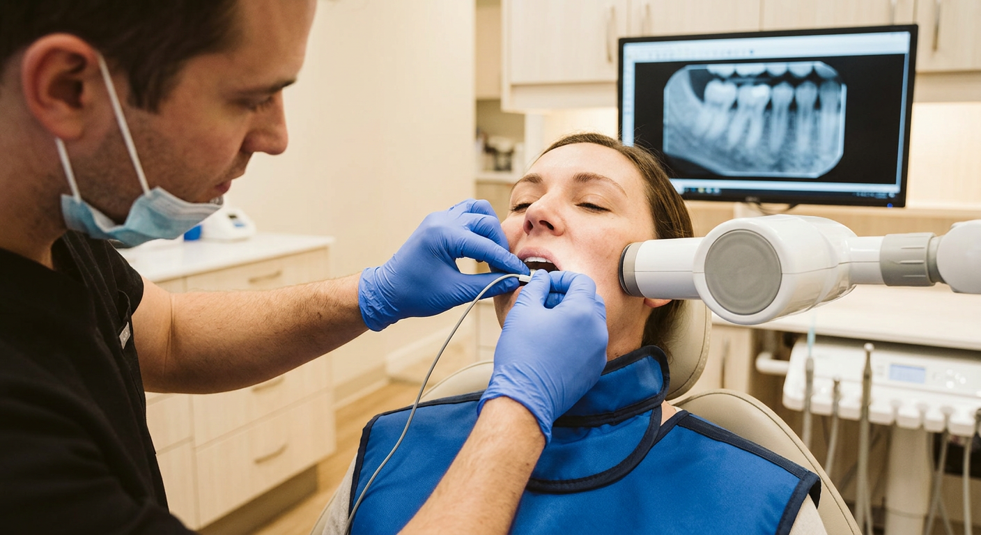
How digital X-rays work: A small sensor or phosphor plate is placed in your mouth, and a brief, low-dose X-ray exposure captures the image. The digital sensor converts the X-ray energy into an electronic signal that’s displayed instantly on a monitor. We can adjust brightness, contrast, and magnification without retaking the image — and share files electronically with specialists when needed.
What digital X-rays help us find: cavities between teeth and under existing fillings, bone loss from gum disease, infections at the root tip (abscesses), impacted or developing teeth, cracks and fractures in roots, and changes in bone density around implants. Regular imaging also helps us track the progression of treatment and catch problems early, when they’re smaller and easier to treat.
Digital sensors require significantly less radiation than traditional film — up to 80% less in many cases. We follow ALARA principles and use the lowest effective dose for every image.
Images appear on-screen in seconds, eliminating darkroom processing delays. This means faster diagnosis, shorter appointments, and the ability to discuss findings with you in real time.
Digital images can be brightened, sharpened, magnified, and color-mapped without retaking the X-ray. This post-processing flexibility helps us detect subtle cavities, hairline cracks, and early bone changes.
We place a small, thin digital sensor in your mouth and position you comfortably. A lead apron and thyroid collar are provided for shielding.
The X-ray exposure lasts just a fraction of a second. The image appears instantly on the chairside monitor.
We review the images together, using magnification and contrast tools to explain any findings — cavities, bone levels, root health, and more.
Images are saved securely in your digital chart. We can compare them to previous X-rays to track changes and share electronically with specialists if needed.
Are digital X-rays safe?
Yes. Digital X-rays use significantly less radiation than traditional film. A set of four bitewing X-rays delivers roughly the same radiation as a few hours of natural background exposure. We follow ALARA guidelines and only take X-rays when clinically necessary.
Do X-rays hurt?
You may feel brief pressure from the sensor against your gums, but the actual exposure is painless and lasts less than a second. If you have a sensitive mouth, let us know — we have smaller sensors and positioning aids to improve comfort.
How often do I need dental X-rays?
It depends on your individual risk. Patients with active decay or gum disease may need bitewings every 6–12 months; low-risk adults may only need them every 18–24 months. We follow ADA and FDA guidelines tailored to your specific history.
Can I have X-rays if I’m pregnant?
Elective X-rays are typically postponed during pregnancy. However, if there’s an urgent dental concern, X-rays can be taken safely with proper shielding. Always let us know if you’re pregnant or may be.
What’s the difference between digital X-rays and CBCT?
Digital X-rays produce 2D images and are used for routine screening (cavities, bone levels). CBCT produces a full 3D volume for complex cases like implant planning, impacted teeth, or pathology. We use each where it’s most appropriate.
Why do you use a lead apron?
The lead apron shields your body from scatter radiation, though the dose from dental X-rays is already very low. It’s a standard safety practice, and we also provide a thyroid collar for additional protection.
Can children have digital X-rays?
Yes, and they’re especially important for monitoring developing teeth, detecting early decay, and evaluating orthodontic timing. We use the smallest sensors and lowest doses appropriate for pediatric patients.
Will I be able to see my X-rays?
Absolutely. We display images on a chairside monitor and walk you through each finding. Understanding what’s happening in your mouth helps you make informed decisions about your care.